Glaucoma Detailed Work-up
Glaucoma is much more common among old people but it happens in any age group as well. Untreated glaucoma can lead to permanent damage of the optic nerve and resultant visual field loss, which over time can progress to blindness.
The nerve damage involves loss of retinal ganglion cells in a characteristic pattern. This can permanently damage vision in the affected eye(s) and lead to blindness. Worldwide, glaucoma is one of the leading cause of blindness. People who are with high myopia, hypertension, cataracts, diabetes, and with immediate family member with glaucoma are at higher risk for glaucoma. Moreover, incorrect usage of steroid will also increase the risk of glaucoma.
Glaucoma can be roughly divided into two main categories, “open-angle” and “closed-angle” glaucoma. Closed-angle glaucoma can be characterized by sudden ocular pain, seeing halos around lights, red eye, very high intraocular pressure, nausea and vomiting. High pressure damages the sensitive optic nerve and results in vision loss, which is an emergency. Open-angle, chronic glaucoma tends to progress at a slower rate and patients may not notice they have lost vision until the disease has progressed significantly. Advanced equipment can help find out glaucoma at early stages. Damage to the optic nerve and impairment of vision from glaucoma is irreversible, so a regular eye examination is strongly advised.
In general, a “normal” pressure range is between 10-22 mm Hg. Optic nerve can also be damaged even though the eye pressure is not very high. It is not yet known why some people’s optic nerves are damaged even though they have almost normal pressure levels. Several painless tests that determine the status of the optic nerve and corneal thickness measurement, and visual fields are used to diagnose glaucoma.
With advanced equipment, we can diagnose patients who already have glaucoma by observing their nerve damage or visual field loss. Optical Coherence Tomography (OCT), can obtain high-resolution images of the anterior segment of the eye and the retina, provide a straightforward method of assessing axonal integrity in multiple sclerosis, as well as macular degeneration. OCT is a non-invasive scanning laser that provides high-resolution images of the retina, helping your Optometrist to evaluate its thickness. OCT can also provide information about the presence and severity of macular edema (swelling).
Our glaucoma detailed work-up includes:
- Review history in general and eye health
- Best visual acuity and refractive status ( lazy eye )
- Stereopsis & binocular vision
- Color vision screening
- Intra-ocular pressure
- Anterior and posterior segment ocular health
- Digital fundus photography
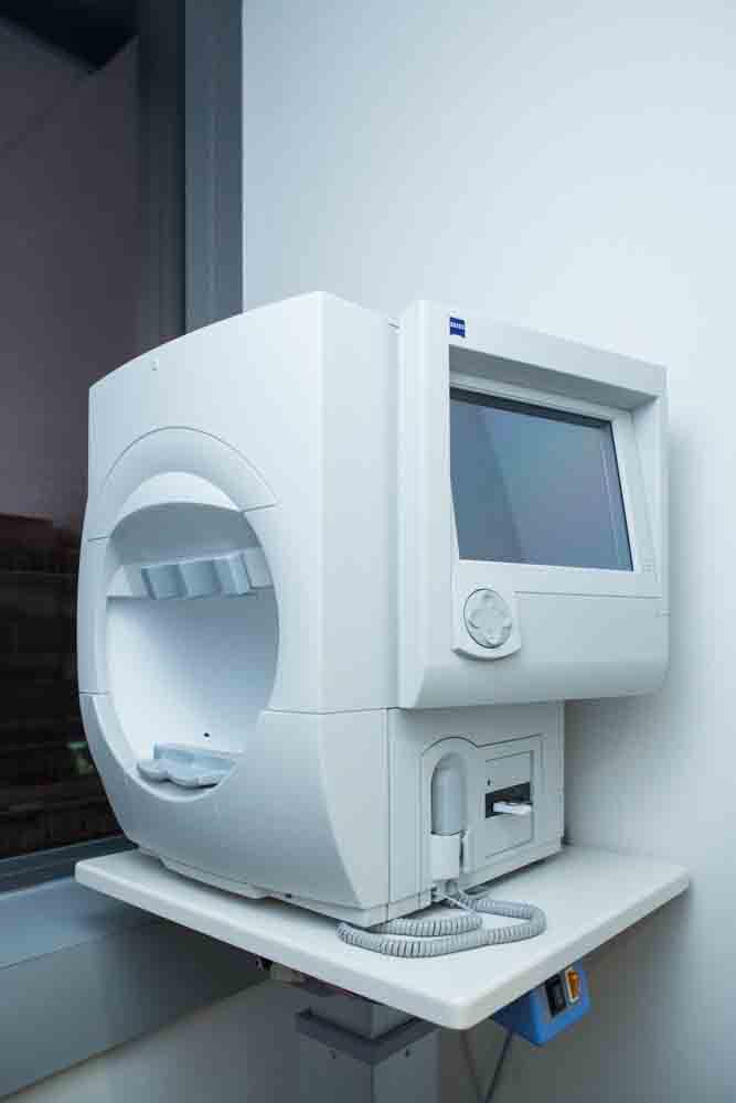
- Visual Field Testing
- Corneal Thickness Measurement
- Optical Coherence Tomography(OCT)
- Diagnoses and treatment plan